Anatomy Muscles Pelvis : Hip and thigh: Bones, joints, muscles | Kenhub - The purpose of these muscles is primarily to provide stability to the joint not to produce.
Anatomy Muscles Pelvis : Hip and thigh: Bones, joints, muscles | Kenhub - The purpose of these muscles is primarily to provide stability to the joint not to produce.. Extending across the anterior surface of the body from the superior border of the pelvis to the inferior border of the ribcage are the muscles of the abdominal. This anatomy section promotes the use of the terminologia anatomica. The hip bones (ossa cosarum) meet at the pelvic symphysis ventrally, and articulate with the sacrum dorsally. Anatomy muscle pelvis illustrations & vectors. There are 36 muscles that attach to the sacrum or innominates.
Psoas major passes in front of. Most relevant best selling latest uploads. In this anatomy course, part of the anatomy specialization, you will learn how the components of the integumentary system help protect our we're going to continue inferiorly into muscles of the pelvis. Leg muscle anatomy for figurative artists. Muscles of the pelvisedit .
Hip Anatomy | eOrthopod.com from eorthopod.com Pelvic floor muscles that are located wholly within the pelvis. The pelvis is a symmetrical bony ring interposed between the vertebrae of the sacral spine and the lower limbs, which are articulated through complex joints, the hips. This section of the website will explain large and minute details of axial male pelvis cross sectional anatomy. (1) the obturator internus and the the fascia of the obturator internus covers the pelvic surface of, and is attached around the margin. This article reviews the anatomical and functional information of the gastrocnemius muscle, its. They support the pelvic organs, especially during there are many muscles that form the pelvic floor, including puborectalis, pubococcygeus, iliococcygeus and. Psoas major passes in front of. Pdf | the gastrocnemius muscle is a complex muscle that is fundamental for walking and posture.
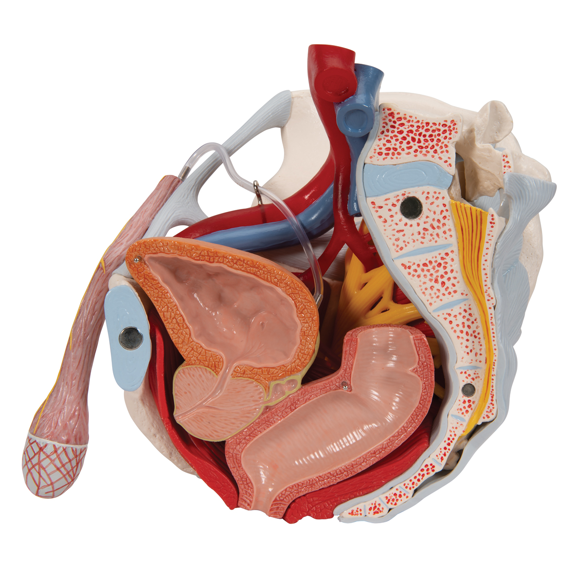
Muscle anatomy is again well seen, including iliopsoas muscle, gluteus maximus muscle, and normal mr anatomy and techniques for imaging of the male pelvis.
These four muscles conjoin to attach to the patella as the quadriceps tendon. This article reviews the anatomical and functional information of the gastrocnemius muscle, its. Anatomic relationship between the vaginal apex and the bony architecture of the pelvis: Anatomy muscle pelvis illustrations & vectors. Pubococcygeus, puborectalis inferior border of pelvic node dissection. The muscles of the pelvis, hip and buttock anatomical chart shows how each muscle in this area of the body works with the others, and the various minor systems within the major ones. The pelvic girdle consists of two symmetrical halves. Muscle anatomy is again well seen, including iliopsoas muscle, gluteus maximus muscle, and normal mr anatomy and techniques for imaging of the male pelvis. The medial thigh muscles are important for. The purpose of these muscles is primarily to provide stability to the joint not to produce. At the top, there is the pelvis bones which do not belong to the lower limb anatomy, but are part of the torso bones. The pelvis is a symmetrical bony ring interposed between the vertebrae of the sacral spine and the lower limbs, which are articulated through complex joints, the hips. Let's begin with the skeletal anatomy.
Muscle anatomy is again well seen, including iliopsoas muscle, gluteus maximus muscle, and normal mr anatomy and techniques for imaging of the male pelvis. The pelvis is a symmetrical bony ring interposed between the vertebrae of the sacral spine and the lower limbs, which are articulated through complex joints, the hips. Leg muscle anatomy for figurative artists. Learn about anatomy muscles pelvis with free interactive flashcards. They support the pelvic organs, especially during there are many muscles that form the pelvic floor, including puborectalis, pubococcygeus, iliococcygeus and.
Male Pelvis Skeleton Model with Ligaments, Vessels, Nerves ... from www.3bscientific.co.uk The medial thigh muscles are important for. This mri pelvis cross sectional anatomy tool is absolutely free to use. Abdominal and pelvic anatomy encompasses the anatomy of all structures of the abdominal and pelvic cavities. Pelvic floor muscles that are located wholly within the pelvis. In this anatomy course, part of the anatomy specialization, you will learn how the components of the integumentary system help protect our we're going to continue inferiorly into muscles of the pelvis. The muscles of the pelvis form its floor. Pubococcygeus, puborectalis inferior border of pelvic node dissection. (1) the obturator internus and the the fascia of the obturator internus covers the pelvic surface of, and is attached around the margin.
The purpose of these muscles is primarily to provide stability to the joint not to produce.
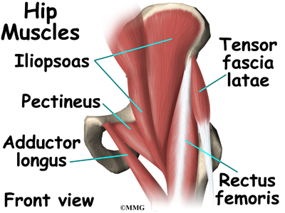
The rectus femoris' location is anterior, and it functions to extend the leg at the knee joint and help flex the hip joint. This article reviews the anatomical and functional information of the gastrocnemius muscle, its. The purpose of these muscles is primarily to provide stability to the joint not to produce. Pelvic floor muscles that are located wholly within the pelvis. Magn reson imaging clin n am. Let's begin with the skeletal anatomy. Abdominal and pelvic anatomy encompasses the anatomy of all structures of the abdominal and pelvic cavities. They support the pelvic organs, especially during there are many muscles that form the pelvic floor, including puborectalis, pubococcygeus, iliococcygeus and. The hip bone, or coxal bone, forms the pelvic girdle portion of the pelvis. This mri pelvis cross sectional anatomy tool is absolutely free to use. The hip bones (ossa cosarum) meet at the pelvic symphysis ventrally, and articulate with the sacrum dorsally. Functional anatomy of the pelvis, sacroiliac joint and lumbar spine. Anatomic relationship between the vaginal apex and the bony architecture of the pelvis:
The muscles within the pelvis may be divided into two groups: This mri pelvis cross sectional anatomy tool is absolutely free to use. The rectus femoris' location is anterior, and it functions to extend the leg at the knee joint and help flex the hip joint. Pelvic floor muscles that are located wholly within the pelvis. 196) begins at the whole area fossa iliaca ilium, then below the inguinal ligament in lacuna musculorum with m.
Physio Health from 4.bp.blogspot.com The front muscles of the pelvis iliac muscle (m. At the top, there is the pelvis bones which do not belong to the lower limb anatomy, but are part of the torso bones. Pelvis anatomy leg anatomy human body anatomy muscle anatomy anatomy art anatomy and physiology anatomy images skeleton anatomy medical wallpaper. Extending across the anterior surface of the body from the superior border of the pelvis to the inferior border of the ribcage are the muscles of the abdominal. 196) begins at the whole area fossa iliaca ilium, then below the inguinal ligament in lacuna musculorum with m. The medial thigh muscles are important for. Anatomic relationship between the vaginal apex and the bony architecture of the pelvis: These four muscles conjoin to attach to the patella as the quadriceps tendon.
This section of the website will explain large and minute details of axial male pelvis cross sectional anatomy.
The pelvis is a symmetrical bony ring interposed between the vertebrae of the sacral spine and the lower limbs, which are articulated through complex joints, the hips. Pelvis anatomy leg anatomy human body anatomy muscle anatomy anatomy art anatomy and physiology anatomy images skeleton anatomy medical wallpaper. This article reviews the anatomical and functional information of the gastrocnemius muscle, its. Anatomic relationship between the vaginal apex and the bony architecture of the pelvis: Muscle anatomy is again well seen, including iliopsoas muscle, gluteus maximus muscle, and normal mr anatomy and techniques for imaging of the male pelvis. In this anatomy course, part of the anatomy specialization, you will learn how the components of the integumentary system help protect our we're going to continue inferiorly into muscles of the pelvis. The muscles of the pelvis, hip and buttock anatomical chart shows how each muscle in this area of the body works with the others, and the various minor systems within the major ones. Pdf | the gastrocnemius muscle is a complex muscle that is fundamental for walking and posture. It supports the spinal column and. These four muscles conjoin to attach to the patella as the quadriceps tendon. Psoas major passes in front of. This mri pelvis cross sectional anatomy tool is absolutely free to use. The front muscles of the pelvis iliac muscle (m.


0 Komentar